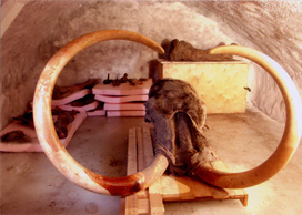
Successful CT measurement and 3D image reconstruction of the Yukagir Mammoth in Japan
- The world's first images of the internal structure of a woolly mammoth in adult size
Its excellent state of preservation and uniqueness make the Yukagir Mammoth a precious specimen of a woolly mammoth. These features ruled out incisions or other destructive procedures as methods of observing its internal parts. For this reason, right from the start, the plans for the program of comprehensive research of the specimen in Japan envisioned the utilization of X-ray computed tomography (CT) equipment to obtain internal anatomical information in the form of image data, without harming the specimen.

This operation was positioned as the central project of the entire program. Under the leadership of Dr. Naoki Suzuki, Vice Chairman of the Scientific Council for Research on Yukagir Mammoth (also Professor of the Jikei University School of Medicine and Director of the Institute for High Dimensional Medical Imaging; the Committee is chaired by Mr. G. V. Tolstych, Minister of Science and Professional Education, Republic of Sakha), the project team succeeded in making CT measurements of the cranium and left front leg, obtaining image data, and reconstructing them into three dimensions. This was the world's first non-destructive acquisition of images of the internal structure of a woolly mammoth in adult size.
1. Course of work
1) Preliminary study
* Advance testing using the skeleton of an Asiatic elephant was conducted in July 2004.
* The team derived CT measurement conditions and determined matters such as the mode of transporting the specimen.
2) Preparation of a capsule for CT measurement
* The preliminary study was followed by the design and manufacture of a capsule for CT measurement of the specimen as is, i.e., in its frozen state, both safely and securely.
* The capsule was made of several kinds of special material, and utilized absolutely no metal. It has a structure enabling not only retention of the specimen in a frozen state for a long time but also biological isolation of its interior from the outside environment.
3) Measurement by a large-scale CT equipment
* The team made measurements with the large-scale CT unit applied for livestock at the National Livestock Breeding Center (in Nishigo-mura, Fukushima Prefecture) from the 17th to the 22nd of December 2004, and obtained basic stratified image data. (It would not have been possible to inspect the specimen cranium, which has a total length of about two meters and weighs more than 250 kilograms, with the ordinary medical CT equipment.)
* The team moved the specimen into the capsule within a refrigerated container, and then carefully placed the capsule into the large-scale CT unit. Measurements were made for about one week.
4) Analysis of image data
* The team took the CT stratified image data to the Institute for High Dimensional Medical Imaging at the Jikei University School of Medicine and commenced analysis. The data consisted of several thousands of pictures.
* The team is reconstructing the data into three-dimensional images upon removal of all kinds of noise occurring during measurement from the series of cross-sectionalimages, and then space combining them and building an image data base for them. This work is still continuing.
2. Results to date
* Among other things, the project revealed the existence of muscular tissue on the face and brain tissue inside the skull, and yielded a substantial amount of anatomical information, as was anticipated.
* Measurement of the volume of the brain upon conversion into three dimensions indicated that it was about the same as that of today's Asian elephant.
* Like those of other elephants, the specimen's cranium contains an intricate bone structure that encloses the brain and contains pockets referred to as “air cells.” In the Yukagir Mammoth, this part was evenly filled with earth. While further study is required, it appears that the specimen sank down below immediately after death, judging from the fairly well-preserved state of the hide and brain tissue.
3. Schedule for the future
* For the future, the study team will continue with its efforts to enhance the image and obtain even more precise 3D images, while also proceeding with the elucidation of minute anatomical structures and physiological functions.
* The results of this research, the 3D images enhanced with computer graphics, and other articles will be on display, along with the frozen specimen, at the Mammoth Laboratory of the Global House throughout the run of EXPO 2005.


